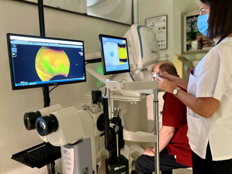
The ophthalmology service of the Torrevieja University Hospital has incorporated a new state-of-the-art diagnostic equipment that allows professionals to observe the fundus through retinography (photography of the retina) without dilating the pupil. It is expected that more than 5,000 patients a year can benefit from this new diagnostic test.
The fundus is commonly explored in ophthalmology, as it allows professionals to detect and monitor many retinal and optic nerve diseases, such as diabetic retinopathy, retinal detachments and tears, age-related macular degeneration, glaucoma, or neuropathies, among others.
Not dilating the pupil is a direct benefit for the patient, avoiding unnecessary discomfort and resuming life normally after the medical examination.
When the professionals had to dilate the pupil to see the fundus, in many cases, the patients had to attend the consultation accompanied since they could not resume their daily chores immediately, nor of course, drive after each examination.
On the other hand, another of the main advantages of the new equipment provided is that it allows for a digital record of the patient’s retina, and thus being able to compare the evolution of diseases over time, with which ophthalmologists can make much more precise decisions.
In addition, as it is a wide-field retinograph, it allows you to see areas of the retina where other conventional retinographs cannot. Therefore, medical professionals can rule out many more peripheral retinal abnormalities.
The scan with the new Optomap equipment usually takes about five minutes. It has no side effects or possible complications, although like all flash photography it can leave some glare for a few minutes.
The team can also perform retinal angiography, which, although it is a test that is used less than in the past, also has its indications.
José Isidro Belda, head of the Ophthalmology service of the Torrevieja health department affirms that “the great advantage of this equipment is to offer better control of retinal and optic nerve diseases in our patients, minimising risks and avoiding unnecessary pupil dilation”.